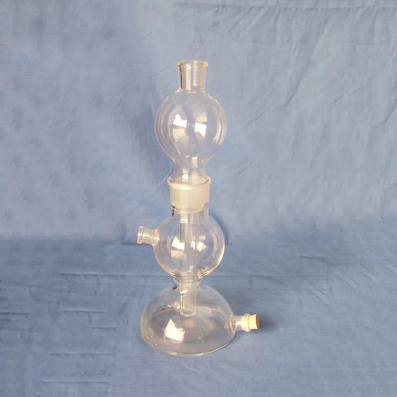
The virus under microscope combines technological innovation and practical design, with distortion-free, clear-quality imaging at every magnification. The mechanical stability and focus precision controls of the virus under microscope ensure accurate specimen positioning. The virus under microscope enhances sample visibility in varying light conditions using a strong illumination system. Optional camera adapters and measuring software are offered to extend its use, making it suitable for various scientific and educational environments.

Applications of the virus under microscope cross into different spheres. It enables disease diagnosis by examining tissue sample and blood smears in medicine. In materials science, the virus under microscope is employed to examine crystal structures, coatings, and composites. In life sciences research, it is used in visualization of cell morphology, patterns of growth, and intracellular action. The virus under microscope also offers quality inspection for production with precision in semiconductor fabrication and microfabrication. It is used in museums and conservation laboratories to examine pigments and fibers in artifacts from ancient times.

The virus under microscope will also evolve by being combined with new quantum and digital technologies. Greater processing speed and improved imaging will capture microscopic motion in real time. Artificial intelligence will decipher complex biological and material structures more accurately than ever before. The virus under microscope will likely consist of interchangeable modular components that can be replaced or reconfigured based on specific research needs. The virus under microscope will remain vital as the scientific frontiers continue to push the frontiers of the unexplored in nature.

Maintenance of the virus under microscope involves regular cleaning and preventive inspection. Always start by making sure all lenses and eyepieces are clean of dust before observing. Avoid subjecting the virus under microscope to extreme temperatures or humidity levels. Clean immersion lenses after each session and remove all the slides from the stage. Keep the virus under microscope covered when not in use to protect it from contaminants. Engage professional maintenance every year to inspect optical alignment and ensure there is smooth mechanical running.
The virus under microscope bridges the visible and invisible by rendering small particles and organisms visible. Using a lens system and controlled light, the virus under microscope enables scientists and students to study samples with utmost precision. It has diverse applications in medicine, biology, electronics, and quality control. Digital and fluorescence forms extend study accuracy, simplifying visualization and data recording in most areas of science.
Q: What are the main parts of a microscope? A: The key components include the eyepiece, objective lenses, stage, focusing knobs, and illumination system, all working together to magnify and clarify specimens. Q: How do you clean the lenses of a microscope? A: Lenses should be cleaned using soft lens paper or microfiber cloth with a small amount of lens cleaner to avoid scratching or damaging optical coatings. Q: What magnification levels can a microscope achieve? A: Depending on the model, a microscope can typically achieve magnifications ranging from 40x to over 1000x for detailed observation of microscopic structures. Q: Why is light adjustment important in a microscope? A: Proper light adjustment ensures accurate contrast and brightness, allowing clear observation without distortion or glare during viewing. Q: Can a microscope be used for educational purposes? A: Yes, microscopes are widely used in classrooms and laboratories to teach students about biology, materials science, and microscopic analysis.
We’ve used this centrifuge for several months now, and it has performed consistently well. The speed control and balance are excellent.
We’ve been using this mri machine for several months, and the image clarity is excellent. It’s reliable and easy for our team to operate.
To protect the privacy of our buyers, only public service email domains like Gmail, Yahoo, and MSN will be displayed. Additionally, only a limited portion of the inquiry content will be shown.
We’re currently sourcing an ultrasound scanner for hospital use. Please send product specification...
We’re looking for a reliable centrifuge for clinical testing. Can you share the technical specific...
E-mail: [email protected]
Tel: +86-731-84176622
+86-731-84136655
Address: Rm.1507,Xinsancheng Plaza. No.58, Renmin Road(E),Changsha,Hunan,China

 af
af
 es
es
 ar
ar
 tr
tr
 sw
sw
 pt
pt
 th
th
 ur
ur
 bn
bn
 ne
ne
 vi
vi
 km
km
 lo
lo
 de
de
 ru
ru
 fi
fi
 nl
nl
 fa
fa
 fr
fr
 ko
ko