Today, clinical laboratories always rely on waters hplc system for the purpose of giving comprehensive chemical and biological data from patient samples. The technology's exceptional sensitivity and accuracy make it possible to separate even the smallest amounts of substances such as drugs and metabolites from complicated mixtures. Laboratory staff performs using waters hplc system in method development, validation and ongoing monitoring of the lab's analytical performance. The multi-use of the instrument guarantees its presence during both normal testing and research work, hence hospitals and laboratories are always consistent in providing accurate and trustworthy diagnostic and analytical results.
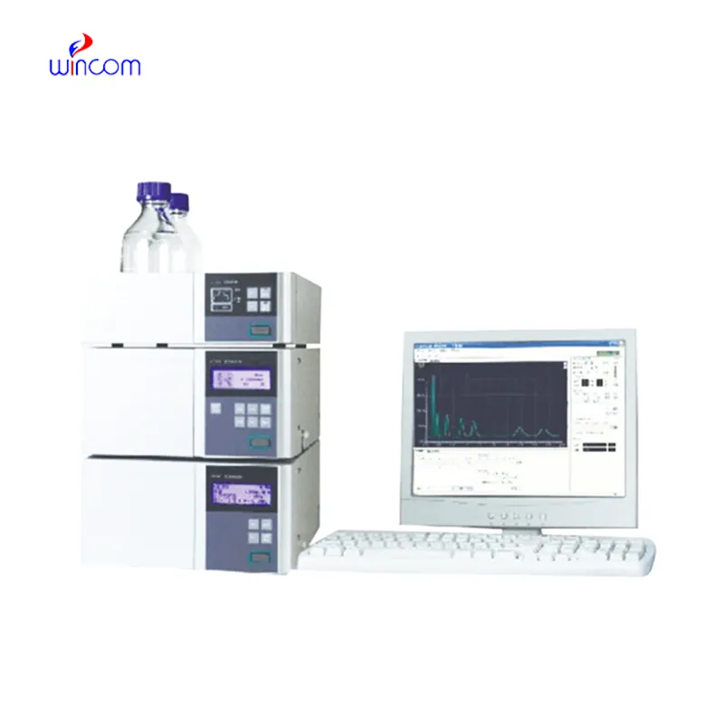
In waters hplc system used to analyze metabolic profiles and biomarkers during clinical research laboratories. It enables the identification of disease markers and monitoring of biochemical changes over time through the separation of small molecules and proteins. waters hplc system also facilitates the study of drug absorption and distribution, toxicity testing, and hospital-based clinical trials and thus making it possible to monitor patient responses to therapies in great detail while at the same time ensuring the accuracy and reliability of the analytical results.
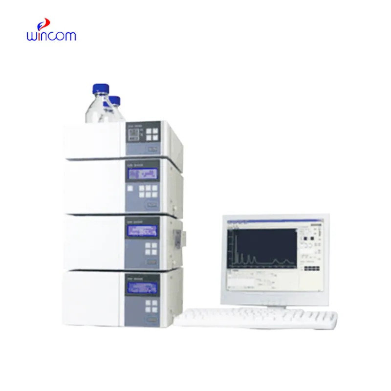
waters hplc system is assigned to become an important player in translational research which is being conducted in hospitals. Among the future developments are the combined detection systems, quicker analysis cycles, and improved reproducibility. waters hplc system will be the mainstay of hospitals' molecular profiling and drug testing along with patient monitoring thus facilitating hospital diagnostics and personalized medicine research.
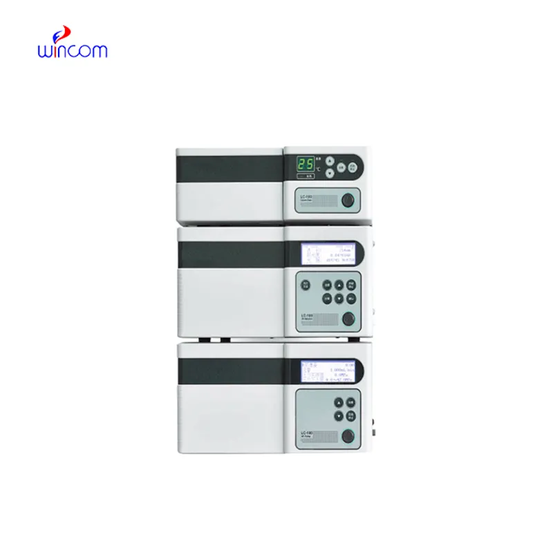
Systematic attention on the system components is necessary for the running of waters hplc system in hospital and research labs. To prevent contamination and pressure problems, flushing of columns, seal replacements, and tubing inspections should be done regularly. Regular calibration of detectors and documentation of maintenance procedures should be done by laboratory technicians. The instruments' life is prolonged by consistent care and monitoring, which also lead to accurate sample analysis and support the reliability of laboratory operations both for clinical and experimental purposes.
The waters hplc system is the backbone of quality control and drug analysis in the pharmaceutical sector. It was able to identify the active ingredients and side products in a very complex, but at the same time, accurate manner. With the choice of proper columns and mobile phases, specialists can isolate the components in both a very efficient and a very constant manner. waters hplc system data is very often requested by regulatory bodies in order to confirm quality of the batch and keep the patients safe. Its accuracy is the mainstay for dosage checking and stability studies. The capability of detecting substances at the trace level renders waters hplc system as the most used and sometimes the only method in drug development, production supervision, and formulation research, thus compliance with industry standards being ensured.
Q: What is HPLC used for in laboratories? A: HPLC turns out to be one of the most significant and essential analytical methods in laboratories equipped with the chemical compound analysis, separation, identification, and quantification of their presence in complex samples which are the research, clinical, and pharmaceutical applications. Q: How does HPLC separate compounds? A: The HPLC separation technique is based on the different affinities of the compounds to the stationary phase and mobile phase within the chromatography column. Q: Can HPLC analyze biological samples? A: Yes, it is certainly possible to carry out analyses on various biological fluids such as blood, serum, urine, etc. for the detection of metabolites, drugs, and biomarkers. Q: How often should HPLC columns be replaced? A: The replacement of the columns must be done according to the manufacturer instructions or when the performance begins to decline, which is quite usual after heavy use or contamination. Q: What detectors can be used with HPLC? A: The analysis type determines the use of, among others, UV, fluorescence, refractive index, and mass spectrometry detectors as the common detectors.
The water bath performs consistently and maintains a stable temperature even during long experiments. It’s reliable and easy to operate.
The centrifuge operates quietly and efficiently. It’s compact but surprisingly powerful, making it perfect for daily lab use.
To protect the privacy of our buyers, only public service email domains like Gmail, Yahoo, and MSN will be displayed. Additionally, only a limited portion of the inquiry content will be shown.
We’re currently sourcing an ultrasound scanner for hospital use. Please send product specification...
Could you share the specifications and price for your hospital bed models? We’re looking for adjus...
E-mail: [email protected]
Tel: +86-731-84176622
+86-731-84136655
Address: Rm.1507,Xinsancheng Plaza. No.58, Renmin Road(E),Changsha,Hunan,China

 af
af
 es
es
 ar
ar
 tr
tr
 sw
sw
 pt
pt
 th
th
 ur
ur
 bn
bn
 ne
ne
 vi
vi
 km
km
 lo
lo
 de
de
 ru
ru
 fi
fi
 nl
nl
 fa
fa
 fr
fr
 ko
ko