The shimadzu x ray machine has been designed keeping in mind the needs of modern healthcare. The system comes equipped with capabilities such as real-time imaging adjustments and intelligent exposure. The digital detector provides high uniformity of images, thus supporting accurate interpretations. The shimadzu x ray machine comes with connectivity solutions that ensure smooth integration with digital radiology networks.

The shimadzu x ray machine plays a central role in preventive medicine since it helps to conduct regular health screenings of the chest, spine, and abdomen. It also identifies early signs of conditions such as osteoporosis and lung disease. The shimadzu x ray machine allows for effective conduct of comprehensive health examinations by physicians.

Future versions of the shimadzu x ray machine will combine energy-efficient technology with high-resolution imaging. Predictive analytics integration will enable early disease detection and personalized screening. Global telemedicine networks will also be enabled by the shimadzu x ray machine, extending access to diagnosis in underserved populations.
Cleaning and calibration are necessary to maintain the shimadzu x ray machine. Surfaces should be cleaned with certified disinfectants to prevent contamination. The shimadzu x ray machine should be tested from time to time to determine performance stability. Electrical grounding and cooling fans also need to be checked periodically to provide safe and efficient service.
The shimadzu x ray machine has been used heavily in various medical fields due to its ability to offer rapid and precise medical images. The shimadzu x ray machine offers precise images of the various body parts that help in the diagnoses of various conditions such as bone injuries, cancer, and infections. The shimadzu x ray machine uses advanced imaging softwares that offer high contrast images.
Q: What types of x-ray machines are available? A: There are several types, including stationary, portable, dental, and fluoroscopy units, each designed for specific diagnostic or operational needs. Q: Can digital x-ray machines store images electronically? A: Yes, digital x-ray machines capture and store images electronically, allowing easy access, sharing, and long-term record management. Q: What safety precautions are required during x-ray imaging? A: Operators use lead barriers, dosimeters, and exposure limit controls to protect both patients and staff from unnecessary radiation. Q: How often should an x-ray machine be inspected? A: It should be inspected at least once or twice a year by certified technicians to ensure compliance with performance and safety standards. Q: Can x-ray machines be used in veterinary clinics? A: Yes, many veterinary clinics use x-ray machines to diagnose fractures, organ conditions, and dental issues in animals.
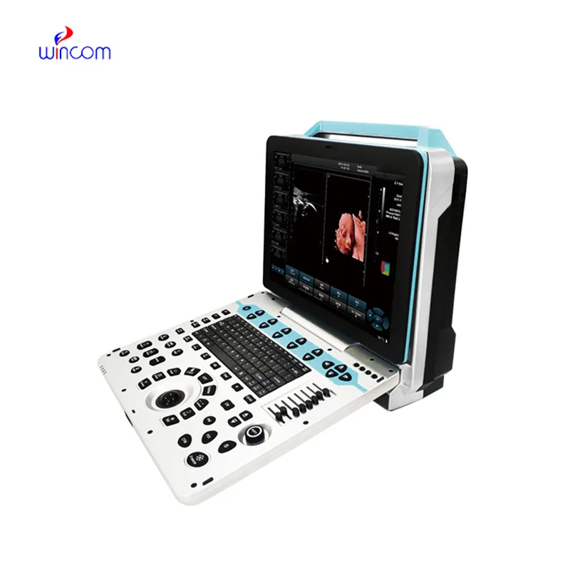
This ultrasound scanner has truly improved our workflow. The image resolution and portability make it a great addition to our clinic.
The centrifuge operates quietly and efficiently. It’s compact but surprisingly powerful, making it perfect for daily lab use.
To protect the privacy of our buyers, only public service email domains like Gmail, Yahoo, and MSN will be displayed. Additionally, only a limited portion of the inquiry content will be shown.
We’re currently sourcing an ultrasound scanner for hospital use. Please send product specification...
We’re interested in your delivery bed for our maternity department. Please send detailed specifica...
E-mail: [email protected]
Tel: +86-731-84176622
+86-731-84136655
Address: Rm.1507,Xinsancheng Plaza. No.58, Renmin Road(E),Changsha,Hunan,China

 af
af
 es
es
 ar
ar
 tr
tr
 sw
sw
 pt
pt
 th
th
 ur
ur
 bn
bn
 ne
ne
 vi
vi
 km
km
 lo
lo
 de
de
 ru
ru
 fi
fi
 nl
nl
 fa
fa
 fr
fr
 ko
ko