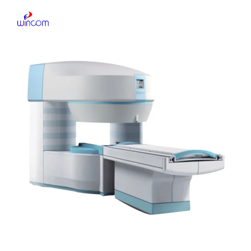
The parts of the x ray machine comes equipped with an intelligent imaging system that increases grayscale depth and detail understanding. The complex algorithms of the parts of the x ray machine improve the viewing of subtle lesions and tissues. The parts of the x ray machine has been designed for high throughput capabilities that promote rapid viewing cycles and convenient data accessibility.

Apart from traditional diagnostics, the parts of the x ray machine is applied in interventional procedures to assist physicians in minimally invasive treatments. It provides real-time imaging during catheter placement, spinal manipulations, and orthopedic implant confirmation. The parts of the x ray machine enhances the accuracy of the procedure and patient safety in complex medical procedures.

Technological progress in the parts of the x ray machine will provide faster image processing, improved 3D visualization, and more accurate diagnostics. Next-generation devices can have AI-assisted positioning systems which will preset imaging settings automatically. The parts of the x ray machine will also be seamlessly integrated into cloud platforms in order to enable instant sharing of information as well as remote consultations.

For the parts of the x ray machine to be trustworthy, maintenance processes must include inclusive system checks, cool-down checks, and cable tests. Preventive maintenance allows potential issues to be noticed early enough before they get worse. The parts of the x ray machine must be monitored for times of use and dates of inspection for traceable records of maintenance.
Through the use of high-tech detectors and digital imaging, the parts of the x ray machine provides high-quality internal structural images. The device enables healthcare providers to track various conditions such as pneumonia, arthritis, and dental cavities. The parts of the x ray machine offers accurate imaging and ease of handling that makes it imperative in diagnostic radiology.
Q: What makes an x-ray machine different from a CT scanner? A: An x-ray machine captures a single 2D image, while a CT scanner takes multiple x-rays from different angles to create 3D cross-sectional views. Q: How is image quality measured in an x-ray machine? A: Image quality depends on factors like contrast, resolution, and exposure settings, which are adjusted based on the target area being examined. Q: What power supply does an x-ray machine require? A: Most x-ray machines operate on high-voltage power systems, typically between 40 to 150 kilovolts, depending on their intended use. Q: Can x-ray machines be used for dental imaging? A: Yes, specialized dental x-ray machines provide detailed images of teeth, jaws, and surrounding structures to support oral health assessments. Q: How does digital imaging improve x-ray efficiency? A: Digital systems allow instant image preview, faster diagnosis, and reduced need for retakes, improving workflow efficiency in clinical environments.
The water bath performs consistently and maintains a stable temperature even during long experiments. It’s reliable and easy to operate.
This ultrasound scanner has truly improved our workflow. The image resolution and portability make it a great addition to our clinic.
To protect the privacy of our buyers, only public service email domains like Gmail, Yahoo, and MSN will be displayed. Additionally, only a limited portion of the inquiry content will be shown.
We’re currently sourcing an ultrasound scanner for hospital use. Please send product specification...
Could you share the specifications and price for your hospital bed models? We’re looking for adjus...
E-mail: [email protected]
Tel: +86-731-84176622
+86-731-84136655
Address: Rm.1507,Xinsancheng Plaza. No.58, Renmin Road(E),Changsha,Hunan,China

 af
af
 es
es
 ar
ar
 tr
tr
 sw
sw
 pt
pt
 th
th
 ur
ur
 bn
bn
 ne
ne
 vi
vi
 km
km
 lo
lo
 de
de
 ru
ru
 fi
fi
 nl
nl
 fa
fa
 fr
fr
 ko
ko