The mini ultrasound scanner combines state-of-the-art digital imaging algorithms that boost contrast and depth perception, which leads to soft tissues being visualized more clearly. The intelligent interface features customizable scanning modes and user profiles for easier operation. Due to its low power consumption and sturdy construction, the mini ultrasound scanner maintains performance that is even in high-demand clinical environments.

In medical imaging, the mini ultrasound scanner is a trustworthy device for different clinical departments. It provides vascular studies support by observing the state of the arteries and veins; in urology, it has a role in the assessment of the bladder and prostate health. The mini ultrasound scanner also enables emergency medical staff to make quick trauma evaluations, directing their actions precisely and efficiently.

The mini ultrasound scanner should integrate with intelligent diagnostic ecosystems and communicate effortlessly with smartphones and electronic records. The synchronized exchange of data in real-time should enable constant patient observation. The next version should focus on improved design, better processing power of artificial intelligence algorithms, and enhanced reconstruction functions.

In order to retain the accuracy of the mini ultrasound scanner, it is important for operators to check the cables and connections of the transducers for evidence of wear. After each use, the surfaces should be wiped clean using non-abrasive cleaners. The mini ultrasound scanner should be turned off properly and covered to prevent dust from collecting. Regular checks by trained personnel should be done.
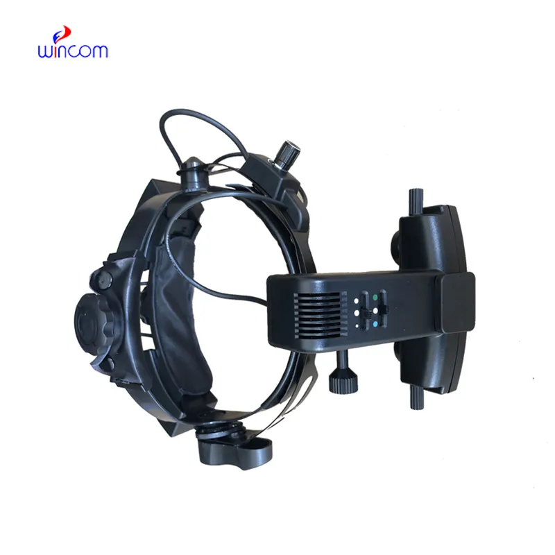
Used in hospitals and clinics, the mini ultrasound scanner provides immediate visual feedback for a variety of medical evaluation uses. Converting sound waves into live images, the mini ultrasound scanner allows physicians to easily detect abnormalities. The mini ultrasound scanner assists with making diagnostic processes safer in addition to improving patient outcomes. It possesses an ergonomic shape alongside digital integration capabilities that support simple data sharing and medical record documentation.
Q: What are the main maintenance requirements for the ultrasound scannert? A: Regular cleaning, proper probe handling, and scheduled inspections help maintain optimal performance. Q: How often should the ultrasound scannert be calibrated? A: Calibration frequency depends on usage levels, but periodic professional checks are recommended. Q: Is the ultrasound scannert suitable for pediatric use? A: Yes, it provides gentle, non-invasive imaging ideal for neonatal and pediatric diagnostics. Q: Does the ultrasound scannert support wireless connectivity? A: Many models include Wi-Fi or Bluetooth features for data sharing and device integration. Q: What materials are used in the ultrasound scannert construction? A: It is built with durable medical-grade components designed to withstand continuous clinical use.
The centrifuge operates quietly and efficiently. It’s compact but surprisingly powerful, making it perfect for daily lab use.
This x-ray machine is reliable and easy to operate. Our technicians appreciate how quickly it processes scans, saving valuable time during busy patient hours.
To protect the privacy of our buyers, only public service email domains like Gmail, Yahoo, and MSN will be displayed. Additionally, only a limited portion of the inquiry content will be shown.
Could you please provide more information about your microscope range? I’d like to know the magnif...
We’re looking for a reliable centrifuge for clinical testing. Can you share the technical specific...
E-mail: [email protected]
Tel: +86-731-84176622
+86-731-84136655
Address: Rm.1507,Xinsancheng Plaza. No.58, Renmin Road(E),Changsha,Hunan,China

 af
af
 es
es
 ar
ar
 tr
tr
 sw
sw
 pt
pt
 th
th
 ur
ur
 bn
bn
 ne
ne
 vi
vi
 km
km
 lo
lo
 de
de
 ru
ru
 fi
fi
 nl
nl
 fa
fa
 fr
fr
 ko
ko