Built from high-quality optics, the microscope electronic eyepiece distributer provides higher clarity for scientific and educational use. The durable body provides stable operation, and the adjustable head and stage setup provide ergonomic convenience. Advanced illumination systems enable observation with high contrast of transparent and reflected samples. The microscope electronic eyepiece distributer is compatible with digital cameras and display devices, enabling real-time observation and recording of microscopic structures for further study and analysis.
Applications of the microscope electronic eyepiece distributer include nanotechnology and public health. In biotechnology, it provides visualization of genes and interactions of cells. In food safety testing, the microscope electronic eyepiece distributer identifies contaminants and microorganisms that affect product quality. In materials engineering, it assists in failure analysis and accurate measurement of microscopic structures. The microscope electronic eyepiece distributer also finds application in archaeology, enabling scientists to study mineral residues and microfossils that reflect environmental conditions in the past.

The future of the microscope electronic eyepiece distributer is influenced by digitalization and smart automation. More efficient imaging sensors will allow the microscope electronic eyepiece distributer to identify three-dimensional structures with unprecedented precision. Artificial intelligence will analyze microscopic images, reduce human errors, and optimize research productivity. Wireless communication and cloud connectivity will facilitate collaboration globally with remote monitoring and immediate data exchange. The microscope electronic eyepiece distributer will be an entirely networked instrument that closes the gap between laboratory precision and data-driven research outcomes.

In the interest of precision and reliability, the microscope electronic eyepiece distributer should be constantly exposed to cleanliness and maintenance. Switch it off at all times before adjusting or cleaning parts. The lenses may be cleaned with alcohol-free cleaners lightly to avoid scratching. Rotary components such as knobs and stage mechanisms value light lubrication at regular intervals. The microscope electronic eyepiece distributer must be stored away from direct sunlight and vibration. Professional checking once a year ensures optical alignment is not affected and prevents wear from invisible damage.
A microscope electronic eyepiece distributer transforms the observation of the unobservable world, revealing patterns, textures, and life beyond the naked eye. It achieves this by illuminating or electronizing a sample by transmitting light or electrons through or above it to produce a magnified image. The microscope electronic eyepiece distributer has widespread uses in science, industry, and education to scan biological tissue, metal surfaces, and nanomaterials. Its ability to unveil subtle details makes it a must-have instrument of observation, measurement, and discovery in modern science.
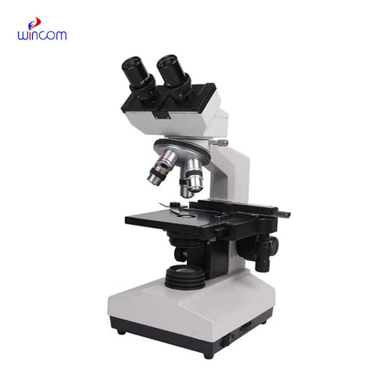
Q: What are the main parts of a microscope? A: The key components include the eyepiece, objective lenses, stage, focusing knobs, and illumination system, all working together to magnify and clarify specimens. Q: How do you clean the lenses of a microscope? A: Lenses should be cleaned using soft lens paper or microfiber cloth with a small amount of lens cleaner to avoid scratching or damaging optical coatings. Q: What magnification levels can a microscope achieve? A: Depending on the model, a microscope can typically achieve magnifications ranging from 40x to over 1000x for detailed observation of microscopic structures. Q: Why is light adjustment important in a microscope? A: Proper light adjustment ensures accurate contrast and brightness, allowing clear observation without distortion or glare during viewing. Q: Can a microscope be used for educational purposes? A: Yes, microscopes are widely used in classrooms and laboratories to teach students about biology, materials science, and microscopic analysis.
The water bath performs consistently and maintains a stable temperature even during long experiments. It’s reliable and easy to operate.
We’ve been using this mri machine for several months, and the image clarity is excellent. It’s reliable and easy for our team to operate.
To protect the privacy of our buyers, only public service email domains like Gmail, Yahoo, and MSN will be displayed. Additionally, only a limited portion of the inquiry content will be shown.
We’re interested in your delivery bed for our maternity department. Please send detailed specifica...
We are planning to upgrade our imaging department and would like more information on your mri machin...
E-mail: [email protected]
Tel: +86-731-84176622
+86-731-84136655
Address: Rm.1507,Xinsancheng Plaza. No.58, Renmin Road(E),Changsha,Hunan,China

 af
af
 es
es
 ar
ar
 tr
tr
 sw
sw
 pt
pt
 th
th
 ur
ur
 bn
bn
 ne
ne
 vi
vi
 km
km
 lo
lo
 de
de
 ru
ru
 fi
fi
 nl
nl
 fa
fa
 fr
fr
 ko
ko