Through a combination of novel imaging software and the inexpensive fetal doppler, the professionals can be very precise with the details of the anatomy they have captured and even more so with the accuracy of the final result. An ergonomic design of the device contributes to the reduction of operator fatigue during long use. The device, the inexpensive fetal doppler, also comes with the ability to connect to more than one probe thus giving it the flexibility required when catering to various diagnostic needs. Besides that, the data export and connection functions of the device help to decrease the time taken in report-generating which is done through image sharing.

The inexpensive fetal doppler is a very significant diagnosis tool used in obstetrics for fetal monitoring and pregnancy development detection. It indirectly affects the cardiology field by providing information about the hearts and blood flow dynamics. Furthermore, the inexpensive fetal doppler is very important in diagnosing abdominal problems, especially issues with the liver, kidneys, and gallbladder. Still, it is also being used in musculoskeletal diagnoses for spotting ligament and tendon injuries.

The future of the inexpensive fetal doppler may include integration with artificial intelligence for automatic analysis of images. Increased portable functionality and wireless communications may improve remote accessibility in healthcare. The inexpensive fetal doppler may include deeper imaging capabilities and rapid data transfer with cloud storage that fosters collaboration on a global medical scale.

Care of the inexpensive fetal doppler involves much more than cleanup. Environmental monitoring and mechanical protection are also part of the process. The inexpensive fetal doppler should not be subject to either vibration or shock. The inexpensive fetal doppler should be backed up periodically to retain vital images.
The inexpensive fetal doppler uses state-of-the-art ultrasound technology to deliver real-time imaging for diagnostic and monitoring purposes. It aids physicians in assessing organs, blood vessels, and soft tissue with unmatched clarity. The non-surgical device is an important tool for guiding medical procedures and making precise diagnoses. The inexpensive fetal doppler combines portability with precision, rendering it extremely useful in routine exams as well as emergency applications.
Q: How does the ultrasound scannert contribute to emergency diagnostics? A: It enables rapid assessment of internal injuries and organ conditions in time-sensitive situations. Q: Can the ultrasound scannert be upgraded with new features? A: Yes, most models support software updates to enhance performance and expand diagnostic functions. Q: What kind of power supply does the ultrasound scannert use? A: It operates on standard AC power and may include rechargeable battery options for mobile use. Q: Is the ultrasound scannert compatible with electronic medical record systems? A: Yes, it can connect to EMR systems to streamline patient data entry and storage. Q: What factors influence the image quality of the ultrasound scannert? A: Image quality depends on probe type, operator technique, and the frequency settings selected for scanning.
The centrifuge operates quietly and efficiently. It’s compact but surprisingly powerful, making it perfect for daily lab use.
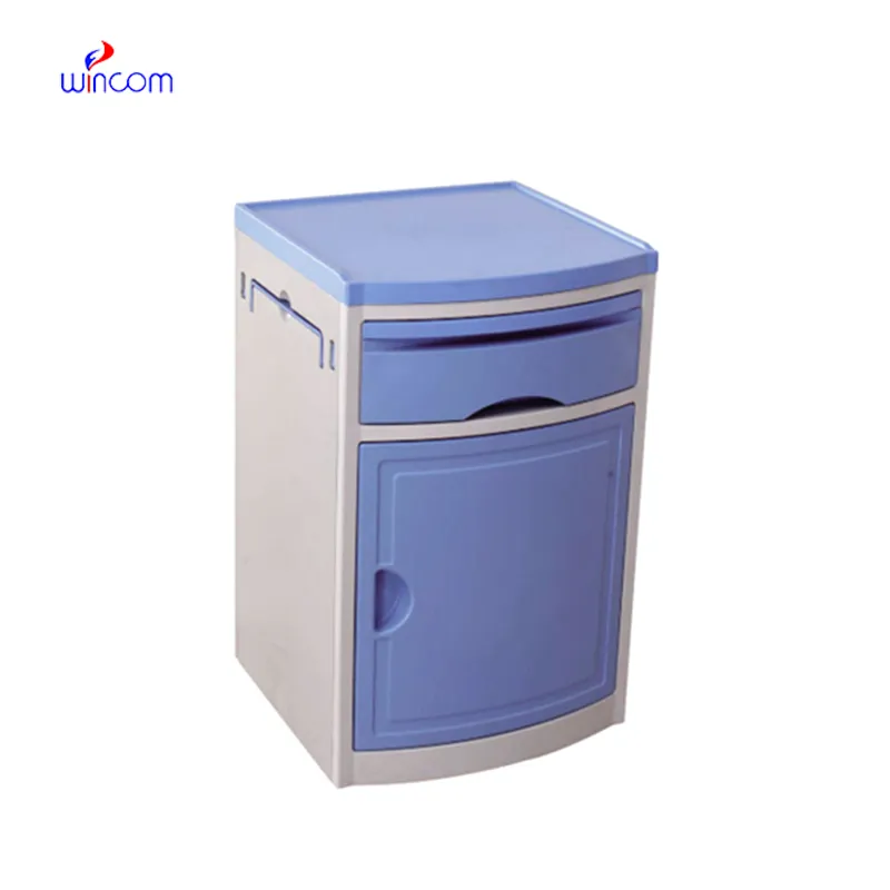
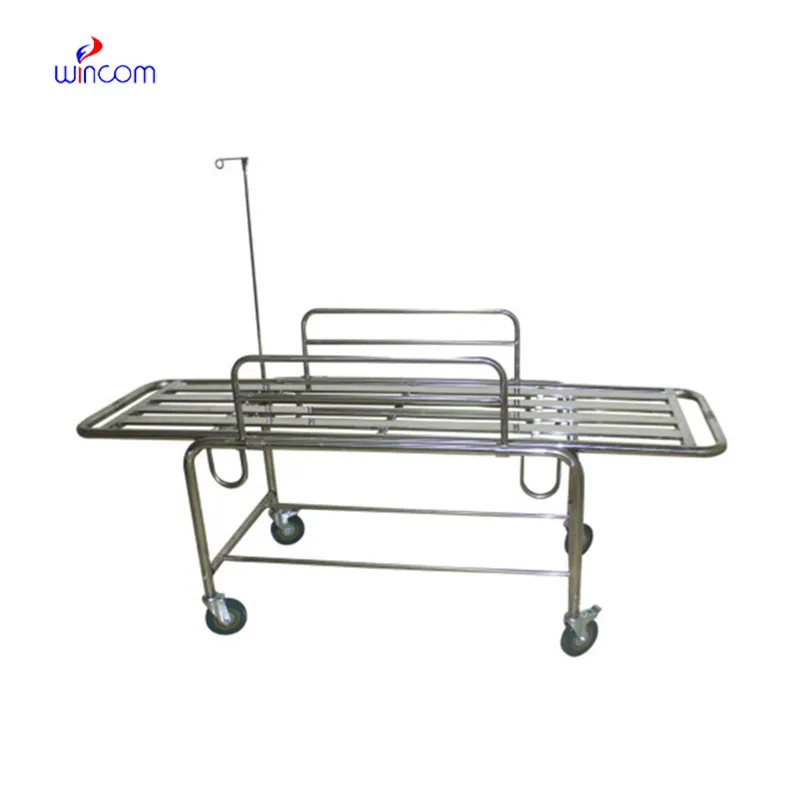
The hospital bed is well-designed and very practical. Patients find it comfortable, and nurses appreciate how simple it is to operate.
To protect the privacy of our buyers, only public service email domains like Gmail, Yahoo, and MSN will be displayed. Additionally, only a limited portion of the inquiry content will be shown.
I’m looking to purchase several microscopes for a research lab. Please let me know the price list ...
Hello, I’m interested in your water bath for laboratory applications. Can you confirm the temperat...
E-mail: [email protected]
Tel: +86-731-84176622
+86-731-84136655
Address: Rm.1507,Xinsancheng Plaza. No.58, Renmin Road(E),Changsha,Hunan,China

 af
af
 es
es
 ar
ar
 tr
tr
 sw
sw
 pt
pt
 th
th
 ur
ur
 bn
bn
 ne
ne
 vi
vi
 km
km
 lo
lo
 de
de
 ru
ru
 fi
fi
 nl
nl
 fa
fa
 fr
fr
 ko
ko