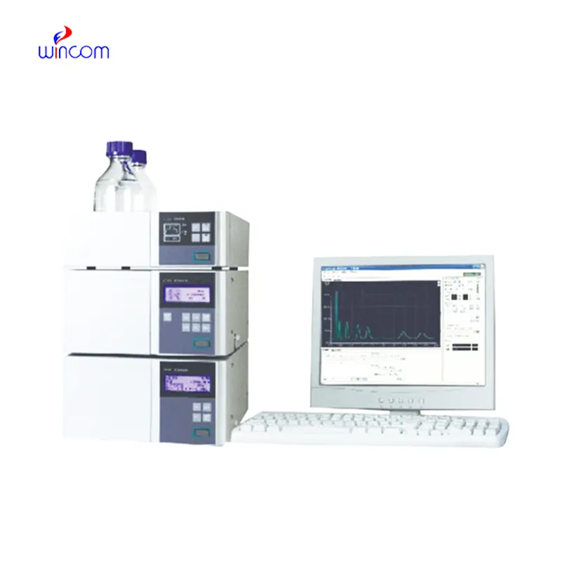
In medical and clinical laboratories, the use of hplc detector results in highly precise determination of therapeutic compounds, metabolites, and biochemical markers. It facilitates creation of detailed patient sample profiles for research and diagnostics. The laboratory personnel prefer hplc detector for confirming method reproducibility, validating analytical procedures, and keeping track of sample integrity. The ultrahigh sensitivity and versatility of the apparatus permit the laboratories to cater to varied applications, thus helping hospitals and research centers to provide reliable and accurate analytical results in various fields of science.
hplc detector is indispensable in the hospital lab for vitamin and nutrient analyses of patient samples. It identifies and determines the amounts of vitamins and minerals that are deficient or excessive in blood or serum. Health care providers depend on it to keep track of patients' nutrition, provide aids for treatment, and assess the impact of supplementation which, thus, boosts the quality of clinical care overall and makes it more beneficial.
The future of hplc detector stresses the integration of hospital information systems and electronic medical records. The analysis of patient samples will be automatically included in the clinical workflows. Increased automation, AI-based interpretation, and better sensitivity will put hplc detector at the center of the laboratory operations and patient care that is focused on the patient's needs.
Systematic attention on the system components is necessary for the running of hplc detector in hospital and research labs. To prevent contamination and pressure problems, flushing of columns, seal replacements, and tubing inspections should be done regularly. Regular calibration of detectors and documentation of maintenance procedures should be done by laboratory technicians. The instruments' life is prolonged by consistent care and monitoring, which also lead to accurate sample analysis and support the reliability of laboratory operations both for clinical and experimental purposes.
hplc detector is equipped with an in-depth examination of biomolecules like proteins, peptides, and nucleic acids. Reversed-phase, ion-exchange, and size-exclusion chromatography methods qualify scientists to get insight into the molecular properties with utmost accuracy. The application of hplc detector in metabolomics studies, enzyme kinetics, and protein characterization helps in high accuracy and reproducibility. The high sensitivity level helps to detect low-molecular-weight molecules in detail and get insight into biological samples at a high level. One of the prime reasons why scientists are interested in hplc detector is its ability to generate information that advances understanding at an advanced biochemistry level.
Q: What is the sample preparation for HPLC? A: For the most part, samples should be filtered, diluted, or subjected to solvent extraction in order to avoid column clogs and have the results be accurate Q: Is HPLC able to pick trace-level compounds? A: With the right detectors, it can pick up such substances in extremely small amounts with high sensitivity. Q: Is HPLC a method that can be applied to analysis of proteins? A: Yes, particularly if one employs size-exclusion and reversed-phase columns for protein, peptide, and biomolecule separation. Q: What is the process of calibrating HPLC? A: The process is done by taking standards of known concentrations that are the same as the one in the sample and using them to check the performance of the column and the accuracy of the detector. Q: Are particular solvents needed for HPLC? A: Yes, the solvents used need to be compatible with the type of the column and the detectors to prevent any damage or interference in the analysis process.
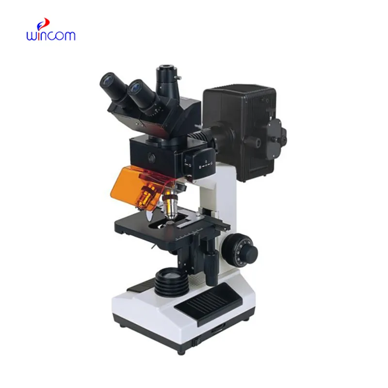
The microscope delivers incredibly sharp images and precise focusing. It’s perfect for both professional lab work and educational use.
The centrifuge operates quietly and efficiently. It’s compact but surprisingly powerful, making it perfect for daily lab use.
To protect the privacy of our buyers, only public service email domains like Gmail, Yahoo, and MSN will be displayed. Additionally, only a limited portion of the inquiry content will be shown.
I’d like to inquire about your x-ray machine models. Could you provide the technical datasheet, wa...
Could you share the specifications and price for your hospital bed models? We’re looking for adjus...
E-mail: [email protected]
Tel: +86-731-84176622
+86-731-84136655
Address: Rm.1507,Xinsancheng Plaza. No.58, Renmin Road(E),Changsha,Hunan,China

 af
af
 es
es
 ar
ar
 tr
tr
 sw
sw
 pt
pt
 th
th
 ur
ur
 bn
bn
 ne
ne
 vi
vi
 km
km
 lo
lo
 de
de
 ru
ru
 fi
fi
 nl
nl
 fa
fa
 fr
fr
 ko
ko