The fetal doppler monitor, being designed for flexibility, brings the user a variety of imaging modes such as B-mode, M-mode, and color Doppler to use. Because of its small size, it can be easily moved from one place to another and that is why it can be used for examining patients in bed. The fetal doppler monitor provides good imaging quality, which can be depended upon in cases of routine diagnostics, fieldwork, and emergency medical interventions.

The fetal doppler monitor has a very wide use in radiology where it supports the guidance of non-invasive procedures. It is very important in gynecology where it is allowed to conduct reproductive system evaluations. In orthopedics, the fetal doppler monitor helps visualize muscles, joints, and tendons ensuring correct diagnostic interpretation. It is this very nature of versatility that makes it a must-have for imaging during procedures in real time.

In future designs of the fetal doppler monitor, eco-efficiency and adaptability factors for the user will be given prominence. Based on advances in probe designs, the system will come equipped with multi-frequency image functionality. The fetal doppler monitor system will also apply predictive analysis capabilities to facilitate early disease detection.

In order to retain the accuracy of the fetal doppler monitor, it is important for operators to check the cables and connections of the transducers for evidence of wear. After each use, the surfaces should be wiped clean using non-abrasive cleaners. The fetal doppler monitor should be turned off properly and covered to prevent dust from collecting. Regular checks by trained personnel should be done.
Used in hospitals and clinics, the fetal doppler monitor provides immediate visual feedback for a variety of medical evaluation uses. Converting sound waves into live images, the fetal doppler monitor allows physicians to easily detect abnormalities. The fetal doppler monitor assists with making diagnostic processes safer in addition to improving patient outcomes. It possesses an ergonomic shape alongside digital integration capabilities that support simple data sharing and medical record documentation.
Q: What imaging modes are available on the ultrasound scannert? A: It supports multiple modes such as B-mode, M-mode, and color Doppler for diverse diagnostic applications. Q: How does the ultrasound scannert improve diagnostic accuracy? A: By providing high-resolution images and real-time feedback, it enables more precise medical evaluations. Q: Can the ultrasound scannert be used in field or remote settings? A: Yes, its portable versions are designed for mobility and can be used in clinics, hospitals, or mobile healthcare units. Q: What kind of display does the ultrasound scannert use? A: It typically features a high-definition digital display that enhances image visualization and readability. Q: How is data from the ultrasound scannert managed? A: The device allows secure storage, easy access, and export of imaging data through USB or network connections.
The hospital bed is well-designed and very practical. Patients find it comfortable, and nurses appreciate how simple it is to operate.
The water bath performs consistently and maintains a stable temperature even during long experiments. It’s reliable and easy to operate.
To protect the privacy of our buyers, only public service email domains like Gmail, Yahoo, and MSN will be displayed. Additionally, only a limited portion of the inquiry content will be shown.
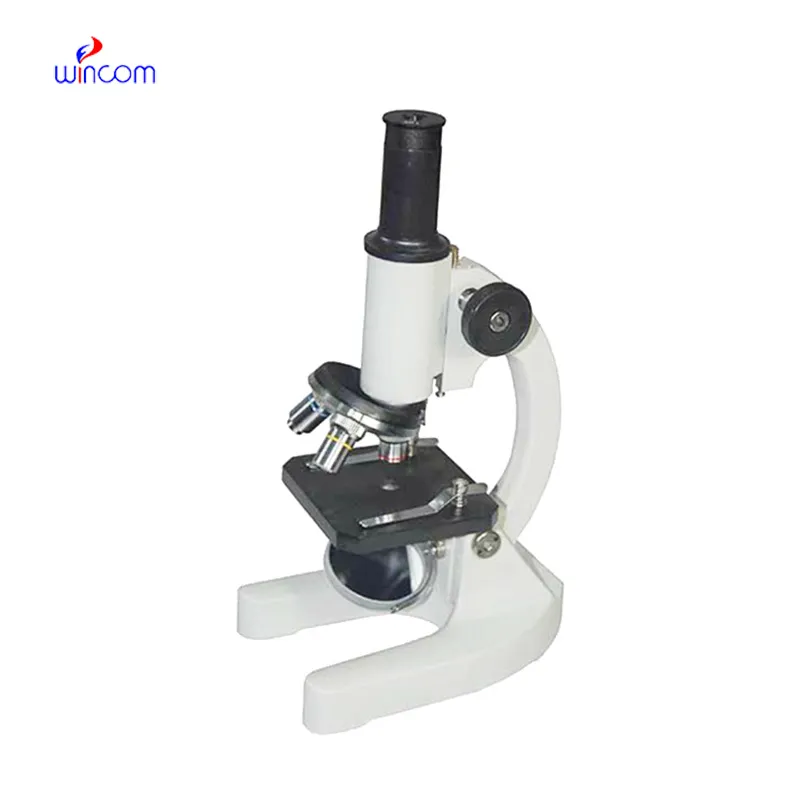
I’m looking to purchase several microscopes for a research lab. Please let me know the price list ...
We are planning to upgrade our imaging department and would like more information on your mri machin...
E-mail: [email protected]
Tel: +86-731-84176622
+86-731-84136655
Address: Rm.1507,Xinsancheng Plaza. No.58, Renmin Road(E),Changsha,Hunan,China

 af
af
 es
es
 ar
ar
 tr
tr
 sw
sw
 pt
pt
 th
th
 ur
ur
 bn
bn
 ne
ne
 vi
vi
 km
km
 lo
lo
 de
de
 ru
ru
 fi
fi
 nl
nl
 fa
fa
 fr
fr
 ko
ko