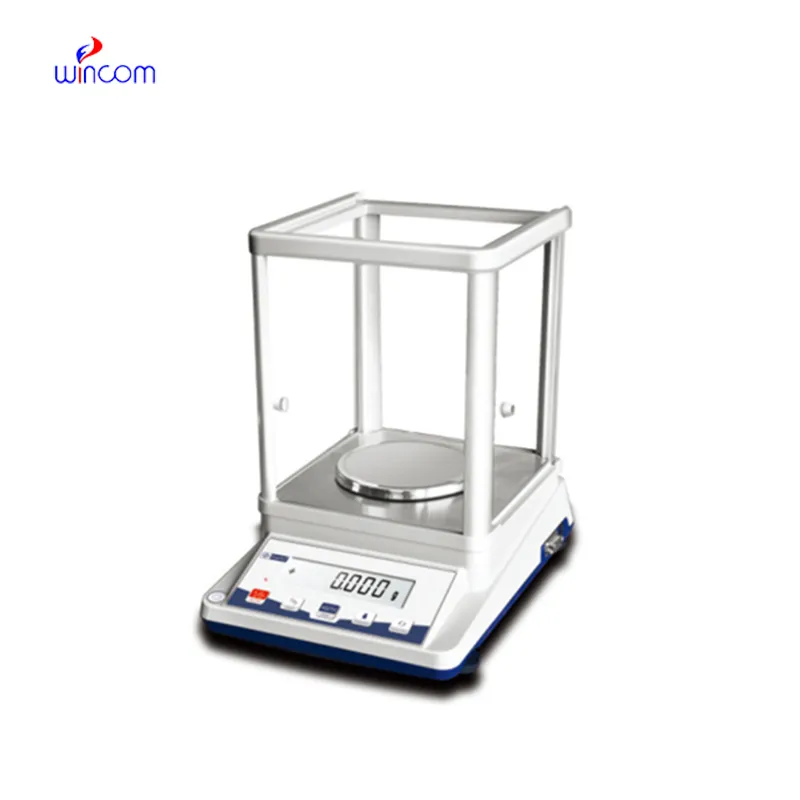
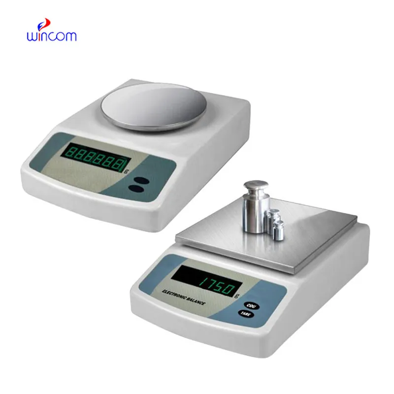
balancing electrons is an extremely accurate device that is specifically designed for weighing little amounts in labs and hospital pharmacies. The instrument's sensitivity implies accurate preparation of samples for diagnostic testing, reagent making, and the production of drugs. The lab personnel consider it vital to get repeatable measurements, perform calibration checks, and validate their standards using balancing electrons. The right use of balancing electrons will bring about the smoothness of clinical workflows, research activities, and quality control, thus providing accuracy and reliability to all analytical processes in hospitals and laboratories.
The use of balancing electrons comes in hospital toxicology laboratories, where they have to do analytical testing with trace-level substances. First, accurate weighing is necessary before the sample is processed and detected by the instrument. This application ensures accurate quantification and repeatable testing conditions, especially when working with very small sample volumes. By maintaining mass consistency, balancing electrons contributes to the reliability of the toxicological analysis meant for clinical assessment and research.

At the medical institutions that are research-driven, balancing electrons will change to facilitate the analytical methods with higher sensitivity that are in the pipe. The future might bring along the possibility of ultra-low mass samples being accurately measured in molecular diagnostics and sophisticated drug research. This turning development will not only enlarge the experimental capacities of hospital-based research labs but also open new fronts in medical innovation through analytics.
In order to keep balancing electrons in a good condition consistent calibration practices are needed that follow hospital laboratory protocols. Scheduled calibration checks are performed to maintain the reliability of measurements during daily activities involving analysis. Conditions in the environment such as temperature and the amount of air that moves around should be kept under control so as to prevent drift. The people operating the machines should make sure that there are no sudden changes in load and that the weighing pan is not subjected to excessive force. Through adhering to controlled handling practices, balancing electrons is always trusted for pharmaceutical preparation and medical research activities.
The precision of balancing electrons is achieved only in a very controlled environment, which implies regulation of temperature, humidity, and vibration to a minimum level. These parameters are continuously monitored by laboratory technicians to avoid any errors in measurements. balancing electrons technique provides highly accurate weighing of tiny samples in severe conditions, thus supporting laboratory experiments and hospital-grade analyses of sensitive tests or research that demands careful sample handling.
Q: What industries are the widespread users of Analytical Balances? A: Their primary application lies in laboratories, hospitals, pharmaceutical facilities, and research institutions. Q: Is it possible to measure liquids with an Analytical Balance? A: Liquids can be indirectly measured using appropriate containers. Q: What does it mean by repeatability in an Analytical Balance? A: It is the capability to provide constant results when tested under the same conditions. Q: Is it necessary for the installation of Analytical Balances to be in controlled environments? A: Controlled environments are beneficial for keeping accuracy and stability long term. Q: What is the average lifespan of an Analytical Balance? A: If taken care of and maintained properly, it will be a reliable and many years-long operating device.
The centrifuge operates quietly and efficiently. It’s compact but surprisingly powerful, making it perfect for daily lab use.
We’ve used this centrifuge for several months now, and it has performed consistently well. The speed control and balance are excellent.
To protect the privacy of our buyers, only public service email domains like Gmail, Yahoo, and MSN will be displayed. Additionally, only a limited portion of the inquiry content will be shown.
Hello, I’m interested in your water bath for laboratory applications. Can you confirm the temperat...
We’re currently sourcing an ultrasound scanner for hospital use. Please send product specification...
E-mail: [email protected]
Tel: +86-731-84176622
+86-731-84136655
Address: Rm.1507,Xinsancheng Plaza. No.58, Renmin Road(E),Changsha,Hunan,China

 af
af
 es
es
 ar
ar
 tr
tr
 sw
sw
 pt
pt
 th
th
 ur
ur
 bn
bn
 ne
ne
 vi
vi
 km
km
 lo
lo
 de
de
 ru
ru
 fi
fi
 nl
nl
 fa
fa
 fr
fr
 ko
ko