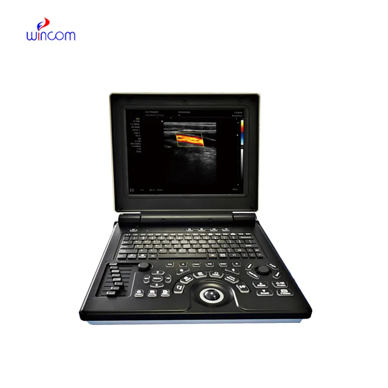
Using state-of-the-art real-time signal processing, the baby fetal doppler has the capability of producing imaging output that is invariably sharp. The system of the device is capable of dynamically tuning the frequency and gain for achieving the best image quality. The baby fetal doppler with its versatile probe compatibility is able to deal with different demanding clinical applications like obstetrics, cardiology, and abdominal scans.

The baby fetal doppler is a tool that medical professionals are using in different departments like pediatrics for the measurement of the size of the organs and neurology for appreciating the anatomy of the soft tissues. It makes it easy for anesthesiologists to perform nerve blocks and vascular access procedures. The baby fetal doppler increase the speed and the trust of the diagnosis during both routine checkups and highly technical interventions.

The coming years will see the evolution of the baby fetal doppler into an independent and adaptable imaging solution. The increased level of automation will eliminate the need for human input. The baby fetal doppler may include predictive model components that will help healthcare providers to identify probable risks to an individual's health.

Proper care and management of the baby fetal doppler needs to be carried out to ensure that it functions well at all times. Cleaning of the probes using a recommended disinfectant helps prevent contamination of the probe and image distortion. Storage of the baby fetal doppler in a clean and dry place away from high temperatures helps prolong the life of the equipment.
Built for performance and accuracy, the baby fetal doppler is an imaging diagnosis platform. It gives real-time images of tissues, organs, and vascular systems, enabling increased detection of pathology. Space-efficient and compact, the baby fetal doppler is ideal for hospitals, clinics, and ambulatory healthcare facilities. Its accuracy imaging enables physicians to offer timely and informed medical care.
Q: What is the primary function of an ultrasound scannert? A: Ultrasound scanners are designed to create real-time images of internal organs, tissues, and blood flow using high-frequency sound waves. Q: How does the ultrasound scannert ensure clear imaging results? A:It uses advanced converter technology and digital processing to enhance image clarity and contrast. Q: In what medical fields is the ultrasound scannert commonly used? A: It is widely used in obstetrics, cardiology, urology, radiology, and emergency medicine. Q: Is the ultrasound scannert safe for repeated use? A: Yes, it is non-invasive and does not emit radiation, making it safe for frequent diagnostic applications. Q: Can the ultrasound scannert store and share imaging data? A: Yes, it supports data storage, retrieval, and digital transfer for easy integration with hospital systems.
We’ve used this centrifuge for several months now, and it has performed consistently well. The speed control and balance are excellent.
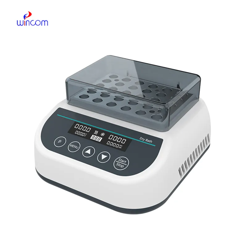
The water bath performs consistently and maintains a stable temperature even during long experiments. It’s reliable and easy to operate.
To protect the privacy of our buyers, only public service email domains like Gmail, Yahoo, and MSN will be displayed. Additionally, only a limited portion of the inquiry content will be shown.
We’re looking for a reliable centrifuge for clinical testing. Can you share the technical specific...
Could you share the specifications and price for your hospital bed models? We’re looking for adjus...
E-mail: [email protected]
Tel: +86-731-84176622
+86-731-84136655
Address: Rm.1507,Xinsancheng Plaza. No.58, Renmin Road(E),Changsha,Hunan,China

 af
af
 es
es
 ar
ar
 tr
tr
 sw
sw
 pt
pt
 th
th
 ur
ur
 bn
bn
 ne
ne
 vi
vi
 km
km
 lo
lo
 de
de
 ru
ru
 fi
fi
 nl
nl
 fa
fa
 fr
fr
 ko
ko