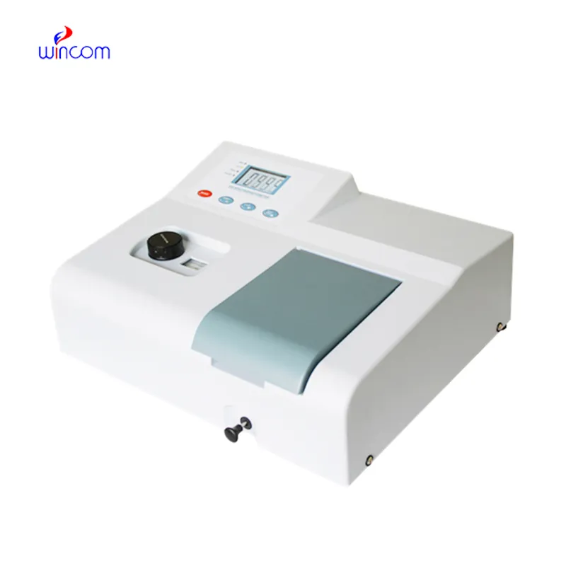
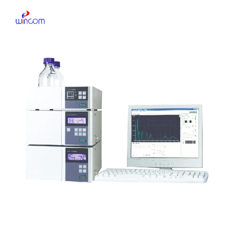
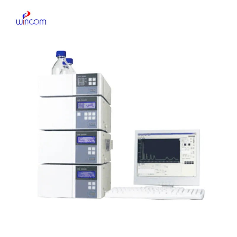
autosampler hplc offers high resolution separation of complex samples in clinical, pharmaceutical, and hospital laboratories, thereby supporting advanced laboratory workflows. It allows performing an in-depth analysis of drugs, metabolites, and small biomolecules. autosampler hplc is used by laboratory staff for research validation, patient monitoring, and method development. Its precision, speed, and adaptability make analytical efficiency greater and at the same time, make consistent and reproducible results which in turn, strengthen laboratory operations in the areas of healthcare and scientific environments.
autosampler hplc allows the personnel of hospitals and laboratories to keep an eye on the presence of environmental pollutants in sterile drugs. It purifies and recognizes the remaining solvents, preservatives, and other possible impurities thus, confirming safety and meeting the requirements of regulatory authorities. This technology is vital in the battle against exposing patients to toxic agents.
Advanced software platforms for predictive analytics in healthcare are going to be part of the autosampler hplc integration. The hospitals will take advantage of the real-time data provided by the patient samples to influence their clinical decisions. Molecular profiling as well as automated quality control and laboratory efficiency will be theautosampler hplc future applications targeting the improvement of patient care.
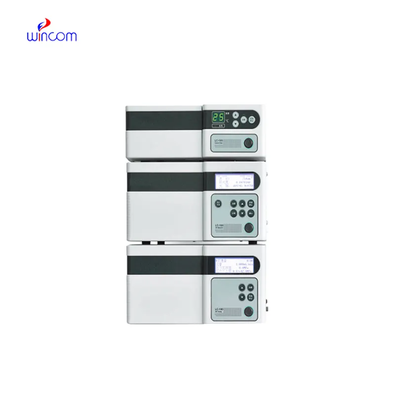
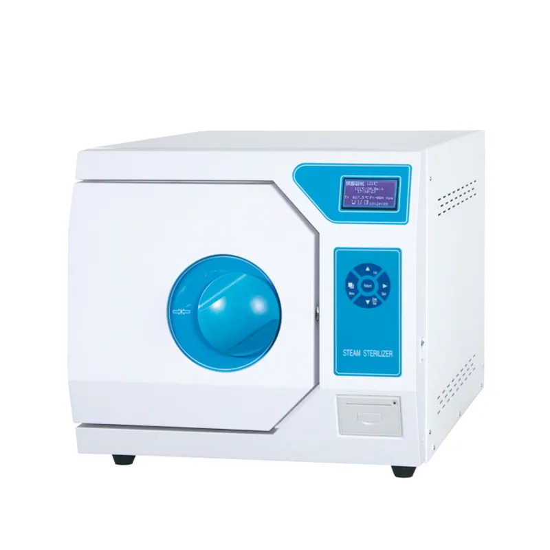
autosampler hplc will require regular maintenance to be kept up in order to continue providing precise measurements in medical laboratories. After every use, the technicians should flush the columns, check the seals, and inspect the tubing for wear and tear and ensure that the detector is working. Regular calibration and good solvent management decrease the chances of system damage and increase the consistency of the results. Good care and maintenance not only increase the efficiency of the laboratory but also help in providing reliable diagnostics and maintaining the instruments for hospital applications.
The autosampler hplc is the backbone of quality control and drug analysis in the pharmaceutical sector. It was able to identify the active ingredients and side products in a very complex, but at the same time, accurate manner. With the choice of proper columns and mobile phases, specialists can isolate the components in both a very efficient and a very constant manner. autosampler hplc data is very often requested by regulatory bodies in order to confirm quality of the batch and keep the patients safe. Its accuracy is the mainstay for dosage checking and stability studies. The capability of detecting substances at the trace level renders autosampler hplc as the most used and sometimes the only method in drug development, production supervision, and formulation research, thus compliance with industry standards being ensured.
Q: What types of HPLC columns are available? A: Reversed-phase, normal-phase, ion-exchange, and size-exclusion columns are the main types of columns used according to the nature of the analytes. Q: Can multiple samples be analyzed simultaneously? A: Yes, in high-throughput systems, automated sample injection and sequential analysis are among the techniques to achieve this. Q: How does temperature affect HPLC performance? A: Temperature changes can cause variations in separation efficiency and retention times; however, the majority of labs make use of precise temperature control. Q: Can HPLC be integrated with data software? A: Sure, it can be linked with laboratory software for data collection, processing, and reporting. Q: What types of laboratories use HPLC? A: HPLC is employed by hospitals, pharmaceuticals, biochemistry research, and environmental testing labs.
This x-ray machine is reliable and easy to operate. Our technicians appreciate how quickly it processes scans, saving valuable time during busy patient hours.
We’ve been using this mri machine for several months, and the image clarity is excellent. It’s reliable and easy for our team to operate.
To protect the privacy of our buyers, only public service email domains like Gmail, Yahoo, and MSN will be displayed. Additionally, only a limited portion of the inquiry content will be shown.
We’re currently sourcing an ultrasound scanner for hospital use. Please send product specification...
Could you share the specifications and price for your hospital bed models? We’re looking for adjus...
E-mail: [email protected]
Tel: +86-731-84176622
+86-731-84136655
Address: Rm.1507,Xinsancheng Plaza. No.58, Renmin Road(E),Changsha,Hunan,China

 af
af
 es
es
 ar
ar
 tr
tr
 sw
sw
 pt
pt
 th
th
 ur
ur
 bn
bn
 ne
ne
 vi
vi
 km
km
 lo
lo
 de
de
 ru
ru
 fi
fi
 nl
nl
 fa
fa
 fr
fr
 ko
ko