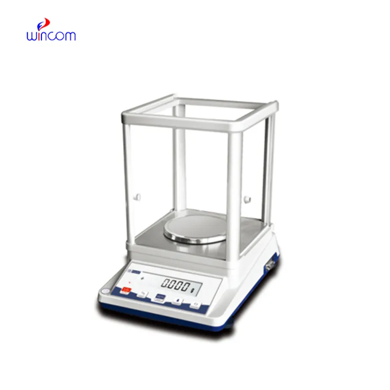
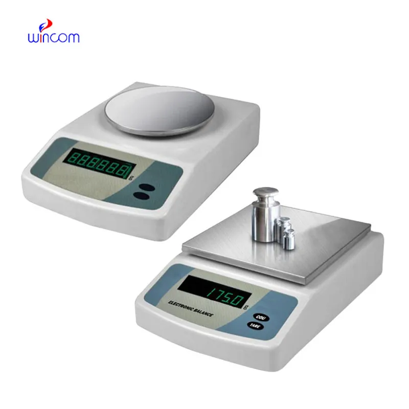
Small samples for accurate mass measurement depend on the laboratory's Weighing Scale that determine the efficiency and reliability. In hospitals, it guides researchers and pharmacy staff in making medicines, calibrators, and experimental solutions. Weighing Scale guarantees very low weight measurement variation which is a precondition for reproducible laboratory results. Its strength, sensitivity, and accuracy are the reasons it is indispensable in clinical laboratories, pharmaceutical testing, and biomedical research where precision is critical.
Hospital research centers utilize Weighing Scale in the process of making experimental medical materials and diagnostic compounds. Accurate mass measurement gives the researchers the power to supervise the formulation ratios during the first evaluation stage. This technology promotes organized experimentation, reproducibility and performance assessment among different research samples. Weighing Scale is making the trustworthiness of experimental workflows in medical innovation environments by supplying the consistent mass data.

The future application of Weighing Scale will be broadened in education laboratories at teaching hospitals. The training provided to lab techs and medical researchers will be accomplished with the help of advanced simulation modes and guided measurement functions. This revolution will offer the medical student the chance to learn practically while the doctor will continue to rely on the precision of the instruments in the lab.
The maintenance of Weighing Scale involves the aspects of storage and inactivity care that come first. The balance should be protected from dust and vibration when it is not in active use. Periodically checking the operational status during long storage prevents unnoticed performance drift. These practices guarantee that Weighing Scale is still capable of accurate use in laboratories, medical and hospital settings.
The precision of Weighing Scale is achieved only in a very controlled environment, which implies regulation of temperature, humidity, and vibration to a minimum level. These parameters are continuously monitored by laboratory technicians to avoid any errors in measurements. Weighing Scale technique provides highly accurate weighing of tiny samples in severe conditions, thus supporting laboratory experiments and hospital-grade analyses of sensitive tests or research that demands careful sample handling.
Q: What maintenance does an Analytical Balance require? A: A periodic cleaning, checking of the calibration, and also verifying the performance are all necessary. Q: Can an Analytical Balance handle continuous daily use? A: Yes, provided that the correct laboratory conditions and rules are followed. Q: Why is leveling important for an Analytical Balance? A: The accuracy and repeatability of the measurements depend on proper leveling. Q: Can Analytical Balances be connected to laboratory systems? A: Most of the models allow connectivity with laboratory information systems. Q: Are Analytical Balances sensitive to vibration? A: Yes, stable weight readings can be disturbed by vibrations.
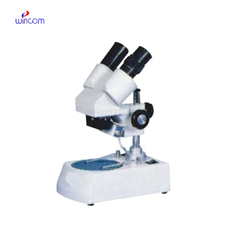
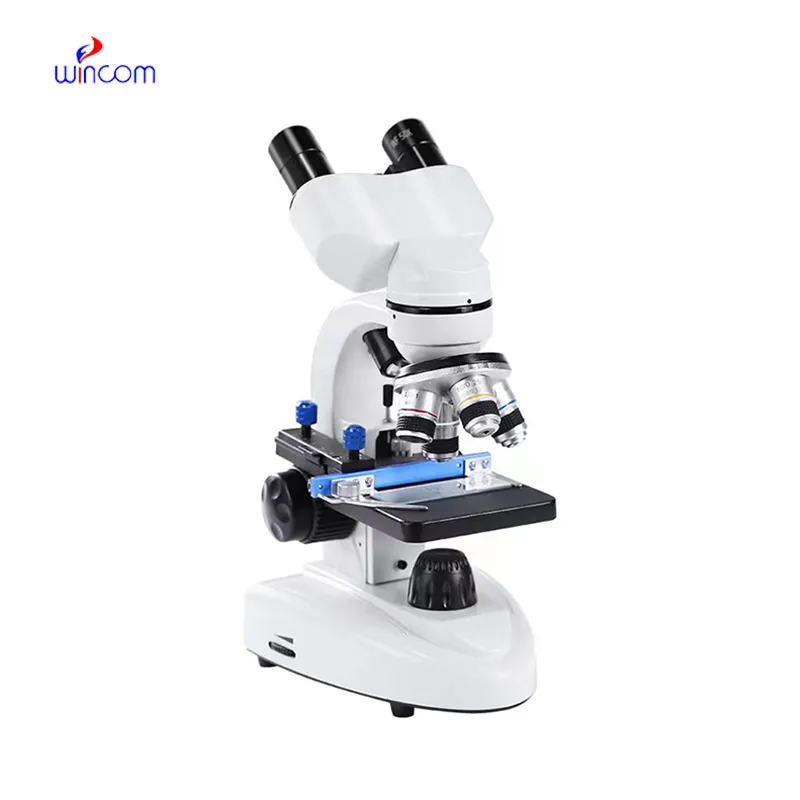
The microscope delivers incredibly sharp images and precise focusing. It’s perfect for both professional lab work and educational use.
We’ve been using this mri machine for several months, and the image clarity is excellent. It’s reliable and easy for our team to operate.
To protect the privacy of our buyers, only public service email domains like Gmail, Yahoo, and MSN will be displayed. Additionally, only a limited portion of the inquiry content will be shown.
We are planning to upgrade our imaging department and would like more information on your mri machin...
I’m looking to purchase several microscopes for a research lab. Please let me know the price list ...
E-mail: [email protected]
Tel: +86-731-84176622
+86-731-84136655
Address: Rm.1507,Xinsancheng Plaza. No.58, Renmin Road(E),Changsha,Hunan,China

 af
af
 es
es
 ar
ar
 tr
tr
 sw
sw
 pt
pt
 th
th
 ur
ur
 bn
bn
 ne
ne
 vi
vi
 km
km
 lo
lo
 de
de
 ru
ru
 fi
fi
 nl
nl
 fa
fa
 fr
fr
 ko
ko