Blog
Eine Krebsdiagnose hängt letztendlich davon ab, dass ein Pathologe einen Objektträger richtig interpretiert. Nichtsdestotrotz beginnt dieser essentielle Schnitt immer mit einem präzisen Schnitt mit einem Mikrotom. Das Rotationsmikrotom ist ein unterschätzter Champion an der entscheidenden Schnittstelle zwischen Präzisionstechnik und klinischer Onkologie.
In diesem Artikel wird untersucht, wie diese lebenswichtige Laborausrüstung vom Labortisch bis zum Krankenbett funktioniert. Wir beschreiben seine grundlegenden mechanischen Prinzipien, bewerten seine diagnostischen und Forschungsanwendungen und fassen wesentliche Kriterien für die Auswahl der Ausrüstung zusammen. Dieser Leitfaden bietet wichtige Informationen für klinische Pathologielabore und Krebsforschungsorganisationen, die ihre Gewebeschnittverfahren verbessern und strategische internationale Kaufentscheidungen treffen möchten.
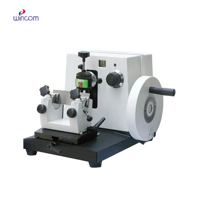
Ein Rotationsmikrotom funktioniert nach einem äußerst präzisen mechanischen Prinzip. Ein Handrad bewegt einen Probenhalter in einem gleichmäßigen, gleichmäßigen Bogen an einer fest angebrachten Klinge vorbei. Bediener haben im Allgemeinen die Möglichkeit, die Schnittdicke von 1 bis 60 Mikrometer (µm) einzustellen. In der Krebshistologie ist die Gewinnung von Schnitten mit einer Größe von weniger als 5 µm unerlässlich, um zelluläre Details und Strukturen effektiv untersuchen zu können. Darüber hinaus müssen Labore sorgfältig zwischen Mikrotomklingen aus Stahl und Wolframcarbid entscheiden. Hochwertige Klingen beeinflussen die diagnostische Klarheit erheblich, indem sie Geweberisse vermeiden und die strukturelle Integrität bewahren.
Labore können zwischen manuellen, halbautomatischen oder vollautomatischen Mikrotomtypen wählen. Jede Wahl bringt einzigartige Kompromisse mit sich:
Anspruchsvolle Modelle verfügen zusätzlich über einen automatischen Probenrückzug. Dieser wesentliche Aspekt der Gewebeerhaltung zieht den Probenblock beim Aufwärtshub leicht zurück und verhindert so, dass die Klinge beim schnellen Schneiden über das Gewebe schleift und es beschädigt.
Der typische histopathologische Prozess besteht aus mehreren strengen, geordneten Phasen. Techniker stabilisieren das Gewebe zunächst in Formalin und umhüllen es anschließend mit einem Paraffinblock (FFPE). Anschließend verwenden sie ein histopathologisches Mikrotom zum Schneiden, fahren dann mit der Färbung und schließlich mit der Objektträgeruntersuchung fort. Das Mikrotom wird genau an der kritischen, unveränderlichen Stelle dieses gesamten Vorgangs positioniert. Techniker sind später im Prozess nicht in der Lage, fehlerhafte Gewebeschnitte zu reparieren.
Bestimmte Krebsarten erfordern eine hervorragende Dünnschnittqualität. Die Diagnose von Brust-, Lungen-, Dickdarm- und Prostatakrebs erfordert präzise Schnittdicken, die normalerweise zwischen 3 und 4 µm liegen. Genauigkeit ist in dieser Situation unerlässlich. Wenn ein Mikrotom Artefakte wie Rattern, Kompression oder Messerspuren erzeugt, können Pathologen die Tumorränder falsch interpretieren oder schädliche Mikrometastasen völlig übersehen.
Neben der grundlegenden Gewebestruktur hängt die heutige Beurteilung einer Krebsbiopsie maßgeblich von hochentwickelten molekularen Tests ab.
Diese anspruchsvollen Tests basieren vollständig auf einer effektiven Antigenspeicherung. Techniker können diese Erhaltung nur mit bemerkenswert sauberen, konsistenten Gewebepräparaten erreichen. Letztendlich erleichtert die Genauigkeit rotierender Mikrotome direkt biomarkergesteuerte Behandlungsentscheidungen, die das Leben der Patienten retten.
Krebsforschungslabore sind auf Rotationsmikrotome angewiesen, um komplexe Krankheitsprozesse zu analysieren und neuartige Behandlungen zu entwickeln. Wissenschaftler untersuchen die Tumormikroumgebung häufig anhand von 3D-Gewebemodellen. Diese eingehende Untersuchung erfordert onkologische Serienschnittmethoden, um die 3D-Struktur von Tumoren präzise nachzubilden. Techniker müssen bei zahlreichen aufeinanderfolgenden Schnitten eine gleichmäßige Schnittdicke beibehalten. Diese Konsistenz ist entscheidend für die Untersuchung von Tumorrändern, die Beurteilung der Angiogenese und die Darstellung der Infiltration von Immunzellen.
Präzises mechanisches Schneiden ist auch für präklinische Gewebeanalysen und Tierstudien von entscheidender Bedeutung. Wissenschaftler nutzen Xenotransplantat-Tumormodelle, um die anfängliche Wirksamkeit von Medikamenten zu beurteilen. Präzise Gewebeschnitte ermöglichen es Forschern, die Infiltration von Medikamenten zu verfolgen und nekrotische Bereiche in der Tumorprobe deutlich zu lokalisieren. Daher benötigen Forschungslabore mit hohem Durchsatz motorisierte Mikrotome mit programmierbaren Schnittvorschubfunktionen, um diese umfangreichen Arbeitsbelastungen effektiv bewältigen zu können.
Darüber hinaus nutzt die Onkologie spezielle Schnittumgebungen für unterschiedliche diagnostische Anforderungen. Während herkömmliche Rotationsmikrotome chemisch fixiertes Gewebe verarbeiten, führen Kryostaten intraoperative Gefrierschnittdiagnosen durch, um ein sofortiges chirurgisches Feedback zu erhalten. Heutzutage betreiben umfassende Krebszentren häufig Hybridlabore. Diese Einrichtungen nutzen sowohl Standard- als auch Gefrierschnittmethoden, um einen vollständigen, schnellen Forschungs- und Diagnose-Workflow zu unterstützen.
Die Beschaffung des richtigen Labormikrotomlieferanten und der richtigen Ausrüstung erfordert eine sorgfältige Bewertung der wichtigsten technischen Spezifikationen. Laborleiter müssen sicherstellen, dass die Maschine den strengen täglichen Anforderungen gerecht wird.
| Technische Spezifikation | Ideale Voraussetzung | Bedeutung in Onkologielabors |
|---|---|---|
| Abschnittsdickenbereich | 1 µm bis 60 µm | Geeignet für Standardhistologieschnitte und spezielle Forschungsschnitte. |
| Mindestinkrement | 0,5 µm oder 1,0 µm | Bietet die ultrafeine Auflösung, die für empfindliche Biopsiegewebe erforderlich ist. |
| Probenorientierung | 8° X/Y-Neigung und 360°-Drehung | Ermöglicht dem Techniker die perfekte Ausrichtung unregelmäßiger Gewebeblöcke mit der Klinge. |
| Klingenkompatibilität | Hohes und niedriges Profil | Stellt sicher, dass das Labor je nach Gewebedichte unterschiedliche Klingen verwenden kann. |
Ergonomie und Sicherheit haben einen wesentlichen Einfluss auf die Effizienz des Labors und die Gesundheit der Mitarbeiter. Hochleistungsdiagnostiklabore profitieren erheblich von Anti-Ermüdungselementen wie ebenen, stabilen Handrädern. Wichtige Sicherheitskomponenten sollten einen zuverlässigen Klingenschutz und einen automatischen Rückzug umfassen, um den Benutzer vor wiederholter Belastung und unbeabsichtigten Schnitten zu schützen. Darüber hinaus sind für Pathologielabore mit begrenztem Platzangebot in Entwicklungsmärkten die Gesamtgröße und das Gewicht der Maschine weiterhin wesentliche Faktoren bei der Kaufentscheidung.
Bei der Suche nach zuverlässigen, hochpräzisen Instrumenten für Ihr Labor sollten Sie hervorragende Optionen wie das in Betracht ziehen Rotationsmikrotom MCT-202 . Dieses Gerät bietet präzise Schnittgenauigkeit, anpassbare Klingenkompatibilität und die ergonomischen Sicherheitselemente, die für anspruchsvolle klinische Umgebungen erforderlich sind, was es zu einer hervorragenden Investition für moderne Pathologieprozesse macht.
Letztendlich müssen globale Einkäufer die Einhaltung strenger Compliance-Standards bestätigen. Beschaffungsagenten müssen ein Mikrotom mit CE-Kennzeichnung, eine ISO 13485-Zertifizierung und gegebenenfalls eine FDA 510(k)-Zulassung anstreben. Ein effektives internationales Servicenetzwerk, das Kundendienst, Zugang zu Ersatzteilen und regelmäßige Kalibrierungsdienste bietet, ist für Labore, die in Entwicklungsmärkten arbeiten, von entscheidender Bedeutung.
Die Zukunft der Krebsdiagnostik hängt maßgeblich von der reibungslosen Integration von Whole-Slide Imaging (WSI) und künstlicher Intelligenz (KI) ab. Digitale Pathologieprozesse erfordern eine äußerst gleichmäßige Schnittdicke für jede einzelne Probe. Diese Anforderung legt einen strengen neuen Standard für die Leistung digitaler Pathologie-Mikrotome fest. Die Wirksamkeit der KI-gesteuerten Tumorerkennung hängt stark von der tatsächlichen Qualität der Eingabefolien ab.
Gleichzeitig beobachten wir einen raschen Anstieg der Nachfrage in den aufstrebenden Onkologiemärkten. Pathologielabore nehmen in ganz Asien, im Nahen Osten und in Afrika rasant zu. Dieses Wachstum stellt für globale Anbieter von Pathologielaborgeräten eine bedeutende Exportchance dar. Darüber hinaus wird die Standardisierung von Mikrotomen als Voraussetzung für internationale Akkreditierungsprogramme für Krebszentren immer wichtiger, einschließlich der Programme, die vom College of American Pathologists (CAP) und der Joint Commission International (JCI) überwacht werden.
Das Rotationsmikrotom ist nicht nur ein zusätzliches Laborgerät; Es ist von grundlegender Bedeutung für die diagnostische Präzision. Vom Forschungslabor bis zum Patientenbett ermöglicht präzises Gewebeschneiden lebensrettende Entdeckungen und wirksame Krebsbehandlungen. Die Investition in ein Premium-Mikrotom stellt grundsätzlich die Zuverlässigkeit Ihres gesamten histopathologischen Prozesses sicher. Wir ermutigen Labore, Gesundheitseinrichtungen und globale Beschaffungsbehörden, noch heute ein Angebot einzuholen oder eine Produktberatung zu vereinbaren, um unsere Auswahl an Rotationsmikrotomen zu entdecken und Ihre Diagnosemöglichkeiten zu verbessern.
Bei einem manuellen Mikrotom muss der Bediener ein Handrad drehen, um das Gewebe voranzutreiben und zu schneiden. Ein motorisiertes Mikrotom nutzt einen internen Motor, um den Schneidhub und den Probenvorschub automatisch anzutreiben. Motorisierte Versionen reduzieren die Ermüdung des Bedieners und sorgen für gleichmäßigere Schnittgeschwindigkeiten, was in Pathologielaboren mit hohem Arbeitsaufkommen von großem Vorteil ist.
Eine Dicke von 3 bis 5 Mikrometern entspricht im Allgemeinen der Dicke einer einzelnen Schicht menschlicher Zellen. Das Schneiden von Gewebe mit genau dieser Dicke verhindert, dass sich Zellen auf dem Objektträger überlappen. Diese Einzelzellschicht ermöglicht es Pathologen, Kern- und Zellstrukturen unter dem Mikroskop klar zu untersuchen, um Krebsanomalien genau zu identifizieren.
Beim automatischen Probenrückzug handelt es sich um eine mechanische Funktion, die den Gewebeblock während des Aufwärtsrückhubs des Mikrotoms leicht zurückzieht (normalerweise etwa 10 bis 50 Mikrometer). Dadurch wird verhindert, dass das Gewebe am Klingenrücken reibt, was sowohl die empfindliche Probe vor Beschädigung schützt als auch die Lebensdauer der Klingenkante verlängert.
Digitale Objektträgerscanner und KI-Diagnosealgorithmen erfordern perfekt flache, gleichmäßige Gewebeproben, um den Fokus beizubehalten und Muster genau zu identifizieren. Falten, Rattern oder ungleichmäßige Dicke, die durch einen schlechten Mikrotomschnitt verursacht werden, können zu digitalen Artefakten führen. Diese Artefakte können KI-Algorithmen verwirren und zu nicht schlüssigen oder ungenauen Diagnoseberichten führen.
Internationale Labore sollten nach Geräten suchen, die über eine ISO 13485-Zertifizierung verfügen, die das Qualitätsmanagement für Medizinprodukte gewährleistet. Achten Sie außerdem auf ein CE-Zeichen (das die Konformität mit europäischen Gesundheits- und Sicherheitsstandards anzeigt) und eine FDA 510(k)-Zulassung, wenn Sie in den klinischen Vorschriften der Vereinigten Staaten tätig sind oder diese standardisieren.
E-Mail: [email protected]
Tel.: +86-731-84176622
+86-731-84136655
Adresse: Rm.1507, Xinsancheng Plaza. Nr. 58, Renmin Road (E), Changsha, Hunan, China


 EN
EN
 af
af
 es
es
 ar
ar
 tr
tr
 sw
sw
 pt
pt
 th
th
 ur
ur
 bn
bn
 ne
ne
 vi
vi
 km
km
 lo
lo
 de
de
 ru
ru
 fi
fi
 nl
nl
 fa
fa
 fr
fr
 ko
ko